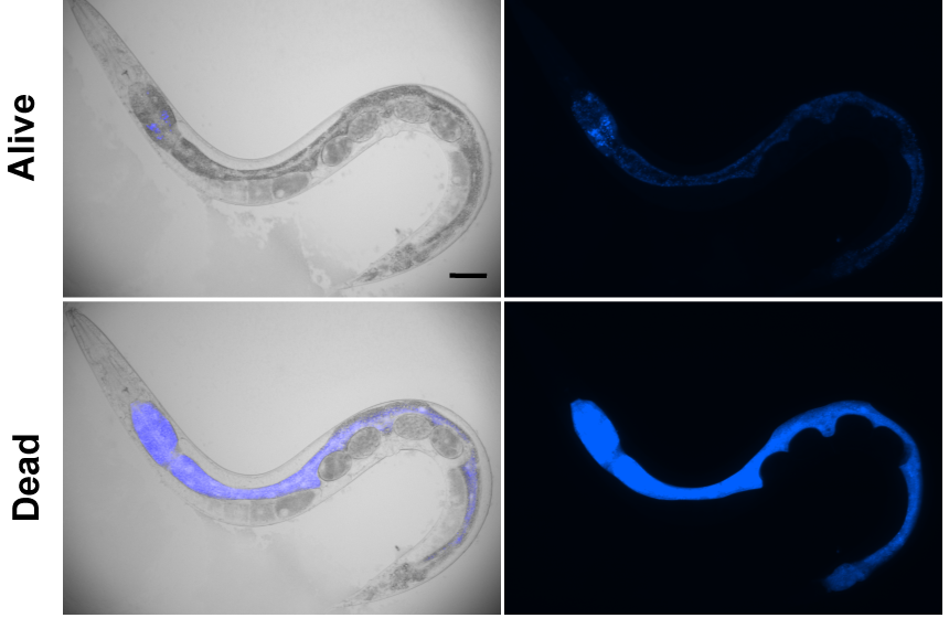
What if we could see the process of death? What would we imagine it to be like? Normally, we are told about seeing a bright light and then… lights out. What if we could document the process of death? What would it reveal to us? What if that bright light was a psychedelic neon blue?
The David Gems’ Lab from the University College London was able to do this elegantly using the nematode worm C. elegans1. These are tiny one millimetre long organisms that are a handy and powerful genetic tool. They are transparent which allows us to quite nicely view their different body components and processes in real time. They are also quite similar to us and a lot of our genes are also conserved in them. They are an excellent model to use for understanding the ageing processes as we can follow them throughout life because they have a lifespan of about 3 weeks2.
In their published work, the David Gems Lab describes the process of organismal death in C. elegans. They describe a new type of blue fluorescence that was observed in dying animals that was previously thought to be linked to lipofuscin which normally accumulates in ageing animals. In their work they detected steady levels of this blue fluorescence in young agile animals and older animals with slow movements. Their findings showed that this blue fluorescence increased by a striking 400% in immobile animals when they were close to death about 2 hrs prior and fades about 6 hours post death1.
This blue wave was not only seen in animals dying of old age but also in stressed induced death (at any age). This stress induced death included freeze-thaw or hot-pick induced killing. They observed that this wave started from the anterior section of the intestine of the animal and flowed towards the posterior and they named the phenomenon Death Fluorescence. The death fluorescence is caused by the production of Anthranilic Acid Glucosyl Esters from the kynurenine pathway. They also showed that the death fluorescence was caused by necrotic cell death. A similar process is also involved in C. elegans neurodegeneration. When there are elevated levels of calcium this leads to uninhibited activation of glutamate receptors resulting in excitotoxicity which causes neuronal cell death1.
This study can give us great insights into the processes that lead to organismal death including how different pathologies (old age, systemic stressed-induced death) lead to mortality and also how certain processes in early life are beneficial but become detrimental when they accumulate with age1.

References
- Coburn C, Allman E, Mahanti P, Benedetto A, Cabreiro F, Pincus Z, Matthijssens F, Araiz C, Mandel A, Vlachos M, Edwards SA, Fischer G, Davidson A, Pryor RE, Stevens A, Slack FJ, Tavernarakis N, Braeckman BP, Schroeder FC, Nehrke K, Gems D. Anthranilate fluorescence marks a calcium-propagated necrotic wave that promotes organismal death in C. elegans. PLoS Biol. 2013 Jul;11(7):e1001613. doi: 10.1371/journal.pbio.1001613. Epub 2013 Jul 23. PMID: 23935448; PMCID: PMC3720247.
- Corsi A.K., Wightman B., and Chalfie M. A Transparent window into biology: A primer on Caenorhabditis elegans (June 18, 2015), WormBook, ed. The C. elegans Research Community, WormBook, doi/10.1895/wormbook.1.177.1, http://www.wormbook.org.
